Scientists using living brain tissue to map human brain
- Dave Lawler, author of Axios World

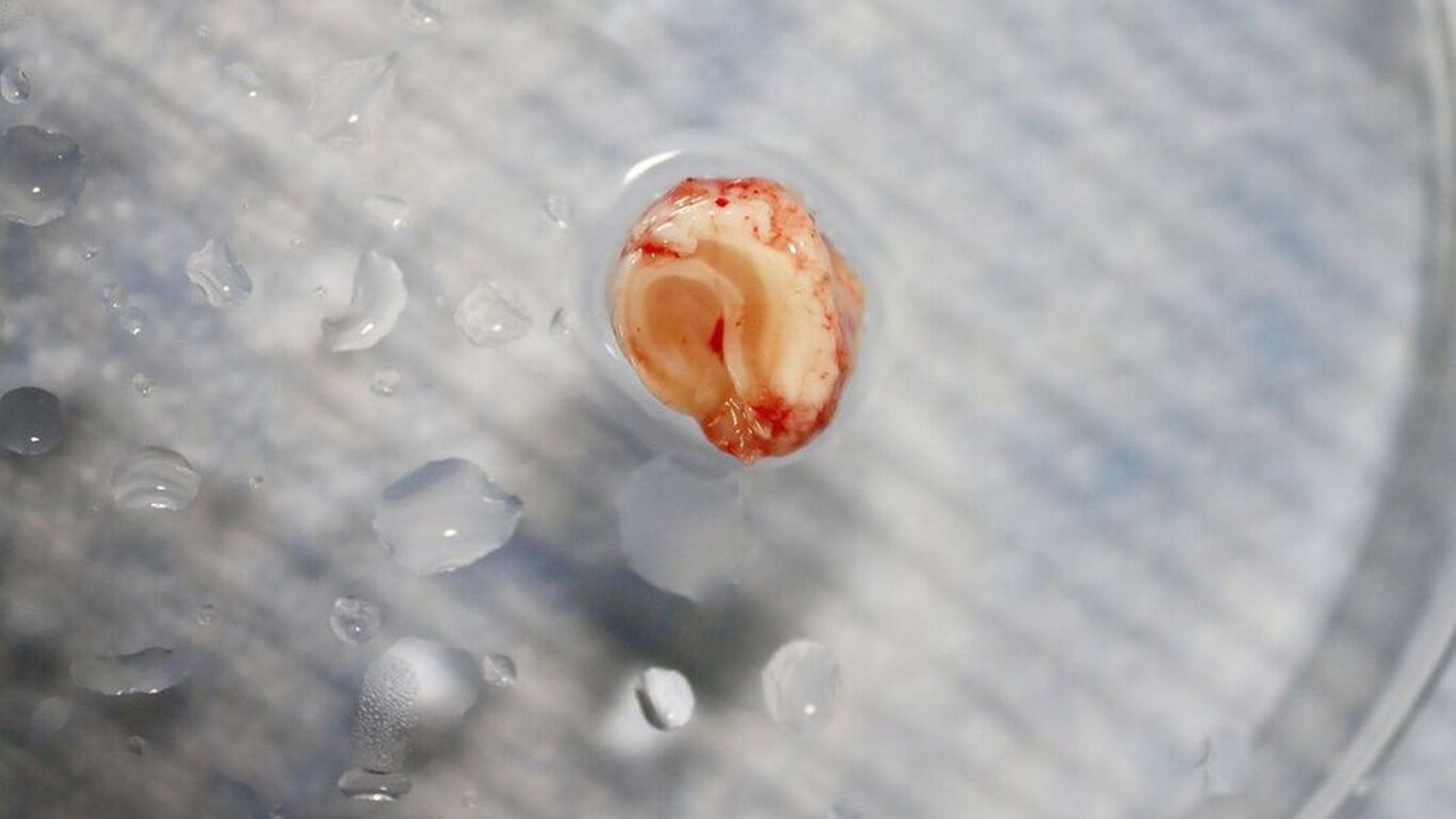
Live brain tissue. Photo courtesy of the Allen Institute.
By studying brain tissue that is typically disposed of during surgery, the Allen Institute for Brain Science in Seattle is building 3D, computer reconstructions of the shape and electrical signals sent by brain cells, per NPR.
Christof Koch, chief scientist and president of the institute, told NPR that previous research on the electrical activity of neurons has relied on samples from animals. This research, therefore, could help indicate what is unique about the human brain and bring better understanding of brain diseases. "In order to understand what makes us who we are, we really need to study the human brain," Koch said.
How they're doing it: During brain surgery, healthy tissue is often removed along with diseased tissue, and then discarded. "So we now have people inside or just outside the operating room who receive those pieces of brain, put them in a special container and then rapidly move them over here," Koch told NPR.
The scientists then keep that tissue alive until they can get them in the lab and study them. So far they've looked at the electrical behavior of about 300 neurons from 36 different people.
The Allen Institute recently received 5-year grants totaling $100 million from the NIH toward its work creating these atlases of brain cells.